The circulatory system distributes the blood that our heart pumps to every organ. They come together to produce the circulatory system. Regular exercise, a healthy, balanced diet, and stress management are all your heart needs to function optimally. But, as you get older, you'll need to incorporate 2D echo test—a vital component of a heart-healthy lifestyle—to check the condition of your heart periodically.
What is 2D Echo Test?
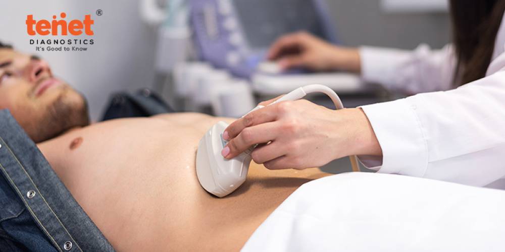
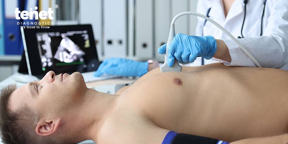
A two-dimensional echocardiography, or 2D echo exam, is a diagnostic imaging process used to assess the anatomy and function of the heart. This non-invasive procedure uses ultrasonic waves to provide real-time, detailed heart pictures. A technician or cardiologist applies a tiny device transducer to the patient's chest during a 2D echo test in Hyderabad. Echoes are produced when high-frequency sound waves from the transducer reverberate off the heart's structures. The ultrasonic pulses have echoes, which are then converted into visual images and shown on a monitor for inspection and analysis. The examination yields essential details regarding the heart's chambers, valves, and surrounding anatomy. It enables medical practitioners to evaluate the heart's dimensions, form, motion, and efficiency of its valves and blood pumping. Additionally, it can reveal whether any illnesses or abnormalities, such as fluid buildup around the heart, congenital heart defects, anomalies of the heart muscle, or problems with the heart valves, are present. Cardiologists frequently utilize the 2D echo test as a diagnostic tool to identify and track various cardiac problems. It usually causes no pain, is noninvasive, and has no severe dangers or adverse consequences for the patient. Trained professionals typically conduct the test at a hospital or specialized imaging center.
Use of 2D Echo Test
To undergo a 2D test, you can search for “2D echo test near me” and visit the nearest diagnostic center. There are various reasons to perform a 2D echo test.
1. Evaluating Heart Function
A 2D echo test aims to assess the heart's composition and operation. It details the heart's chambers and valves' dimensions, form, and function. This aids in evaluating the heart's blood pumping efficiency, figuring out the heart's muscle mass, and spotting any irregularities in the heart's operation.
2. Diagnosing Heart Conditions
A 2D echo test is frequently employed to identify a range of cardiac ailments. It can locate anomalies, including congenital heart deformities, pericardial illnesses (inflammation or fluid accumulation around the heart), heart muscle anomalies (e.g., cardiomyopathy), and heart valve disorders (e.g., valve stenosis or regurgitation). Accurate diagnosis and treatment planning are facilitated by the test's ability to pinpoint the precise location and kind of cardiac issue.
3. Monitoring Heart Disease Progression
For those with established heart diseases, routine 2D echo tests may be carried out to track the disease's development and evaluate treatment efficacy. Doctors can monitor changes in the heart's size, structure, or function over time and use this information to modify treatments, drugs, or surgical alternatives as necessary.
4. Preoperative Evaluation
A 2D echo test may be carried out to assess the patient's heart's anatomy and function before several heart procedures or interventions. This aids in the planning of the surgery and evaluating the risks involved by the surgical team.
5. Follow-up After Treatment
A 2D echo test may be performed following heart surgery, such as valve replacement or repair, or intervention, such as angioplasty, to gauge the overall improvement in heart function and the procedure's success.
6. Screening
A 2D echo test may be performed in some circumstances, particularly in those who have a family history of heart problems or who have risk factors for heart disease. It can assist in recognizing the first indications of cardiac issues, allowing for prompt action and avoiding consequences.
Procedure of 2D Echo Test
About one hour is needed for the 2D Echo test. Any jewelry made of metal will need to be taken off. Following that, your chest will have tiny adhesive electrodes affixed to it. An ECG machine is linked to the electrodes. A transducer will be inserted into your chest and moved around to capture an image of your heart after a gel has been applied to it. A transducer is placed on the chest wall during a 2D Echo test, and the transducer transmits ultrasonic waves reflected by the heart structures. The waves are subsequently processed to produce a real-time image of the heart on a monitor. The patient must lie on their left side for the procedure, which usually takes thirty minutes. The technician could urge the patient to hold their breath or shift postures to obtain various views of the heart. The average 2D echo test price in Hyderabad ranges from Rs. 500/- to Rs. 1500/- depending on various factors.
Different Types of Echocardiograms
Echocardiography comes in various varieties. Each has unique advantages for identifying and treating heart disease. Among them are:
1. Transthoracic Echocardiogram
An ultrasound exam that creates images of your heart using sound waves is called a transthoracic echocardiogram, or TTE. TTE can assess the health of your heart and pinpoint the root causes of symptoms related to the core. Following the test, which is either noninvasive or minimally invasive, you can get back to your regular activities right away.
2. Transesophageal Echocardiogram
A transesophageal echocardiogram, or TEE, is an echo test that makes images of your heart using sound waves. Unlike other echo tests, a TEE produces images from inside your body. The medical professional inserts a flexible, thin tube down your esophagus. Numerous issues, such as blood clots and heart infections, can be diagnosed by a TEE.
3. Stress Echo Cardiogram
An ultrasonography of your heart before and after activity is called an exercise stress echocardiogram. It generates dynamic visuals that demonstrate how effectively your heart works under pressure. This test is used by providers to identify or track cardiac issues and make sure you receive the necessary care.
Final Words
To sum up, the echocardiogram, also known as the 2D Echo doppler test, is a crucial diagnostic instrument in cardiology that offers important insights into the composition and operation of the heart. This noninvasive technique allows medical personnel to evaluate cardiac problems using sound waves to build a detailed heart image. A thorough view of the heart from many perspectives is provided by the several 2D Echo test types, such as transthoracic and transesophageal echocardiograms, which are designed to meet specific diagnostic demands. The process's versatility is reflected in the range of applications it can be used for, including routine cardiac exams, congenital heart defect identification, and cardiac function monitoring during surgical operations.
Frequently Asked Questions
1. How is 2d echo test done?
A transducer will be inserted on your chest and moved around to capture an image of your heart after a gel has been applied to it.
2. Is fasting required for 2d echo test?
No, fasting is not required in order to perform a routine 2D echo test. It is a totally non-invasive process that doesn't require any samples of blood or urine.
3. What is a 2d echo with doppler test?
The heart's blood flow is measured via a Doppler echocardiography, which also determines its direction and speed.
4. Why 2d echo test is done in pregnancy?
It is done to see the structure and function of an unborn child's heart. It is usually done in the second trimester, between 18 to 24 weeks.
5. Which is better, stress test or 2d echo?
Since the stress echocardiography directly visualizes the anatomy and function of the heart during stress, it is more accurate than the stress electrocardiogram (ECG) in identifying coronary artery disease and other cardiac problems.