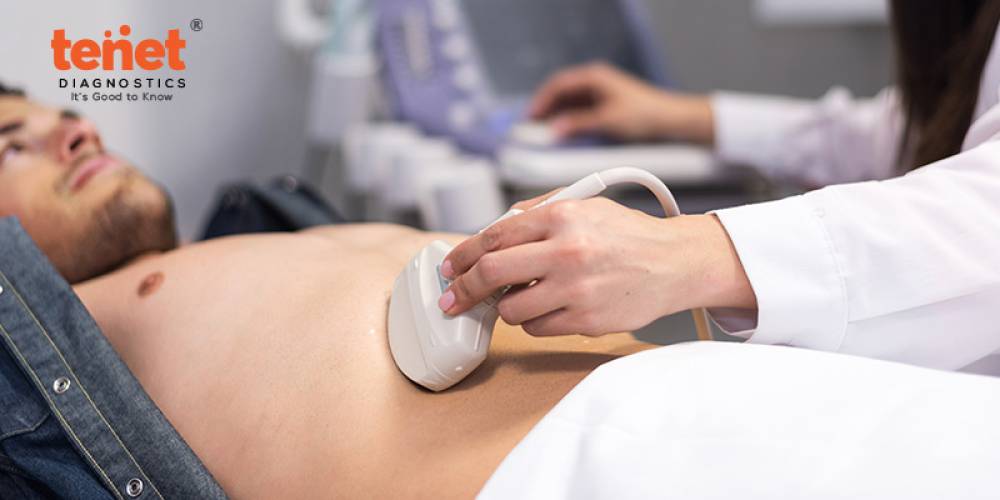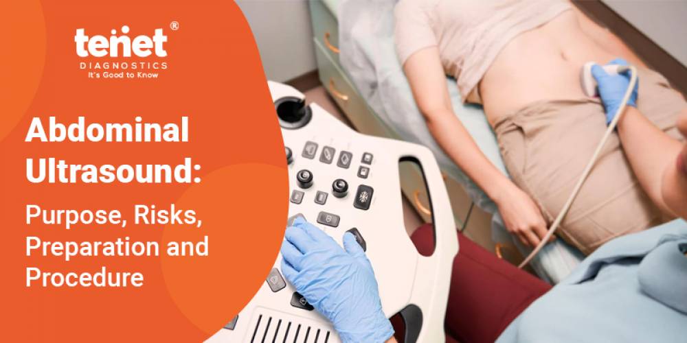
An abdominal ultrasound is a non-surgical procedure used to evaluate the organs and tissues of the stomach. The liver, gallbladder, kidneys, bile ducts, and abdominal aorta are all included. Using ultrasound technology enables fast imaging of abdominal organs and structures outside the body. Blood flow to abdominal organs can also be assessed using ultrasound.
Ultrasonic employs a transducer, which emits ultrasound waves at a frequency that is too high to be heard. The ultrasound transducer is applied to the skin, and the ultrasonic waves travel through the body to the organs and structures. The sound waves will echo off the organs and return to the transducer. The reflected waves are processed by the transducer and then turned into an image of the organs or tissues studied by a computer.
Sound waves will travel at varying speeds depending on the type of tissue met, with bone tissue being the quickest and air being the slowest. The transducer translates the rate at which sound waves are returned to the transducer and how much of the sound wave returns as distinct tissue types. Let's read more about what an abdominal ultrasound is for, the risks, and how it works.
Purpose of Abdominal Ultrasound

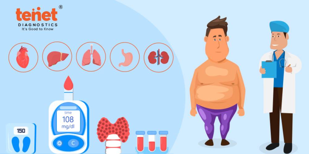
The abdomen contains several main organs. Images of these organs can be created using abdominal ultrasonography. This can aid in identifying stomach disorders that may produce symptoms.
· The Appendix
· Large And Small Intestines
· Stomach
· Gallbladder
· Liver
· Pancreas
· Bladder
· Spleen
are all major organs in the abdomen.
In addition to the organs, ultrasonography can be used to examine other critical structures in the stomach, such as tissues, blood arteries, and abnormal growths. A doctor, for example, may want to examine the abdominal aorta, which transports blood between the heart and the abdominal organs.
In some circumstances, a Doppler ultrasonography may be required. Changes in blood flow through the abdomen can be detected with ultrasound.
Because the abdomen houses so many vital organs, various disorders might arise. These issues might range from inflammation to aberrant hormone reactions, and they can occasionally signal the existence of another ailment.
Use an Abdominal Ultrasound to diagnose problems such as:
· Stomachache
· Liver Related Problems
· Kidneys Stones
· Gallstones
· Inflammatory Disorders: Appendicitis, Pancreatitis, And Abdominal Aortic Aneurysm
· Tumors And Other Abdominal Growths
· Pyloric Stenosis
· Hernia
· Cancer
Abdominal Ultrasound Risks
Abdominal ultrasound has no concerns or risks. Ultrasounds, unlike X-rays and CT scans, do not employ radiation. Therefore, it is recommended to be used by pregnant women to monitor the development of their newborns. In addition, fetal ultrasound imaging provides real pictures of the fetus. Although there is little risk, ultrasound scans should be performed only if there is an apparent medical reason. This is done to avoid exposing the fetus to unnecessary ultrasounds.
Ultrasound imaging and heartbeat monitors are not harmful to fetuses. However, ultrasonography can gently heat tissues in the belly, causing tiny air bubbles in some tissues in some circumstances. The long-term consequences of this are unknown.
Abdominal Ultrasound Preparation

They are fasting for eight to twelve hours before your ultrasound is required. This happens because undigested food may block sound waves, making it harder for the technician to acquire a clear picture.
You must eat a fat-free meal the day before your gallbladder, liver, pancreas, or spleen examination and fast until the operation. You must, however, drink water and take any drugs prescribed by your doctor.
To achieve better-visualized images, you should drink many glasses of water at the time starting up to the test and control your urine to ensure that your bladder is full.
Before the examination, inform your physician about any prescription drugs, under medications, or herbal products you are taking.
Abdominal Ultrasound Procedure
Let us read about how to do an abdominal ultrasound test. Try comfortable clothing that is easy to remove or partially remove if necessary. Sometimes, you may be required to remove your clothing or wear a robe.
However, the ultrasound technician may frequently access the body part being scanned without removing your garments.
The technician will have to apply a water-based gel to the area so that the transducer can smoothly slide over your skin with no air. The gel also aids in the transmission of sound waves. While the test is running, any specific markers will be detected, and measures will be taken.
A typical ultrasound lasts 30 minutes to an hour. You should not feel any discomfort throughout the exam, and you should remain awake and alert throughout. During the abdominal ultrasound test, a technician will often discuss what they are seeing, but in some cases, you may need to wait to discuss the results with your doctor.
A transducer, comparable to a microphone, emits a high-frequency sound that is not detectable to human ears. When the waves strike a dense item, such as an organ or bone, they reverberate. These echoes are later returned to a computer. The echoes are then recorded to evaluate soft tissues and organs' size, shape, and consistency. Finally, this data is transmitted to generate visuals on a computer screen. Ultrasound technicians or sonographers who have received special training in the method usually perform the test. Based on the area being inspected, you may need to shift postures to allow the technician better results.
A radiologist will interpret the ultrasound images. This technology can aid in the diagnosis and treatment of certain illnesses. After the process, the gel will be removed from your skin, and you can resume your normal activities.
Certain factors or diseases may interfere with ultrasonography readings. These could include:
Food in the stomach, leftover barium in the intestines from a recent barium operation, severe obesity. In some tests, you ingest barium, which allows your doctor to visualize your stomach and gastrointestinal tract.
Follow-Up After Abdominal Ultrasound Test

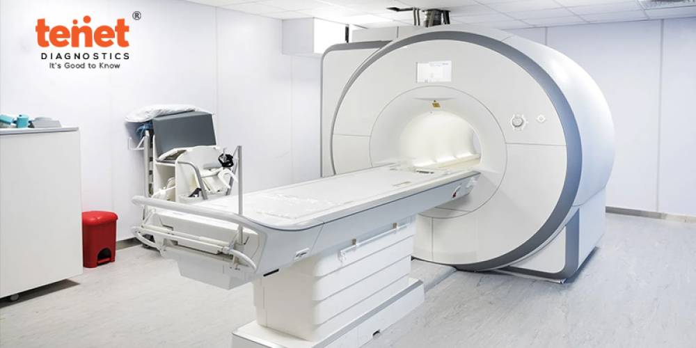
After the physical exam, your physician will review the images and search for unusual characteristics. You will be updated with the results when they are available. If your physician can diagnose your condition based on the results of your abdominal ultrasound, treatment could start almost immediately. However, suppose an abnormality is found that cannot be detected with ultrasound alone. In that case, you may be required to undergo additional diagnostic procedures such as a CT scan, an MRI, or a biopsy sample of tissue, depending on the area that is being investigated.
What Is the Cost of Abdominal Ultrasound? The Abdominal Ultrasound test price in Hyderabad is around INR 1100. You can get the best ultrasound test in Hyderabad by searching for the "abdominal ultrasound near me."
Final Words
Providers frequently use abdominal ultrasonography to diagnose routine and emergency health issues. Unlike several other imaging examinations, ultrasound does not involve radiation. Abdominal ultrasonography is used by doctors to monitor the health of mothers and babies throughout pregnancy. Certain circumstances (such as an empty or full stomach) can cause ultrasound images to be hazy or less detailed. To ensure accurate results, always adhere to any test preparation instructions provided by your provider.