An X-ray is an imaging test that utilizes a small amount of radiation to create images of the body's organs, tissues, and bones. When focused on the chest, it can detect anomalies or disorders of the airways, blood vessels, bones, heart, and lungs. X-rays of the chest can also reveal whether you have fluid in your lungs or if there is fluid or air surrounding your lungs.
The most popular radiological investigation in the emergency department is a chest x-ray 1. The chest x ray PA view is commonly used to help diagnose various acute and chronic diseases involving all thoracic cavity organs. It also serves as the most sensitive plain radiograph for detecting free intraperitoneal gas. It also finds pneumoperitoneum in patients with acute abdominal pain.
If you visit your doctor or the emergency room with chest pain, a chest injury, or shortness of breath, you will most likely be advised for a chest X-ray. Your doctor can use the image to see if you have heart problems, a collapsed lung, pneumonia, fractured ribs, emphysema, cancer, or any of a number of other disorders.
A normal chest X ray is a simple, rapid, and effective examination used to view some of your most essential organs for decades. Some people undergo a series of chest X-rays to see if a health problem improves or worsens.
Why is a Chest X-Ray Done?
X-rays of the chest are a popular method of examination. An X-ray of your chest is used to see how you're reacting to treatment. If your doctor suspects heart or lung illness, a chest X-ray is usually one of the first tests you'll have.
A chest X-ray discloses many things inside your body, including:
Heart-Related Lung Problems
Chest X-rays sometimes reveal changes or issues in your lungs due to cardiac problems. Congestive heart failure, for example, can cause fluid in the lungs.
The Condition of Your Lungs
Chest X-rays can identify cancer, infection, or air collecting in the area around a lung, which can cause the lung to collapse. They can also show chronic lung diseases like emphysema and cystic fibrosis and the difficulties that come with them.
The Size and Outline of the Heart
Heart changes in size and shape can indicate heart failure, fluid surrounding the heart, or difficulties with the heart valves.
Blood Vessels
Because the outlines of the main vessels around the heart — the aorta, pulmonary arteries, and veins — can be seen on X-rays. They can indicate aortic aneurysms, other blood vessel issues, or congenital heart disease.
Postoperative Changes
Chest X-rays help monitor your recovery after having surgery on your heart, lungs, or esophagus in your chest. After surgery, your doctor can examine any lines or tubes to look for air leaks and fluid or air buildup regions.
Calcium Deposits.
Chest X-rays can reveal the presence of calcium in your heart or blood vessels. Its presence could indicate the presence of lipids and other substances in your vessels and damage to heart valves, coronary arteries, heart muscle, or the heart's protective sac. Calcified nodules in the lungs are usually the result of a formerly cured infection.
Fractures.
Chest X-ray helps identify Rib or spine fractures or other problems with the bone.
A Pacemaker, Defibrillator, or Catheter
Pacemakers and defibrillators have wires attached to the heart to help control your heart rate and rhythm. Catheters are tiny tubes that are used to deliver drugs or dialysis fluid. After such medical devices are implanted, a chest X-ray is routinely conducted to ensure that everything is in place.
Why do I need a chest X-ray?
If your doctor suspects that the symptoms are related to chest problems, they may request a chest X-ray. Signs to look out for include:
• Chest Pain
• Persistent Cough
• Fever
• Shortness of Breath
Risks
If you get chest X-rays frequently, you may be concerned about radiation exposure. However, the amount of radiation emitted by a chest X-ray is minimal — even less than that emitted by natural radiation sources.
Even if the benefits of an X-ray exceed the risks, if you need several scans, you may be provided a protective apron. The treatment can be done so that the radiation does not reach your abdomen. If you think you could be pregnant, tell your doctor.
How to Prepare for a Chest X-Ray?
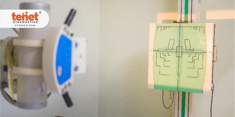
The X-ray is done in a specific chamber with a movable X-ray camera on a long metal arm. You will stand next to a "plate," which could contain X-ray film or a special sensor that records the images on a computer.
The X-ray technician will instruct you on how to stand and will acquire front and side chest views. While the photos are being taken, you must hold your breath so that your chest remains still. If you move, the visuals may become distorted. Denser elements, such as bone and heart muscles, will appear white as the radiation passes through your body and onto the plate.
X-rays are generally painless procedures. As the radiation goes through your body, you are unaffected. You can have the exam while seated or lying down if you have difficulty standing.
Your job is done once the photographs have been captured, which should take about 20 minutes. You can put your clothes back on and go about your day.
Results
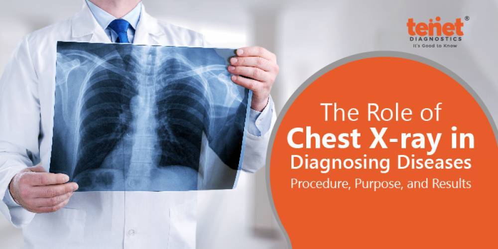
A chest X-ray gives a black-and-white image of your inside organs. Structures that block radiation seem white, while those that allow it to pass appear black.
Because your bones are so dense, they appear white. A lighter region appears around your heart. Because your lungs are full of air and block very little radiation, they appear on the scans as darker spots.
The radiologist, a specialist who has been trained to interpret X-rays and other imaging exams — evaluates the images for signs that could indicate heart failure, fluid around the heart, malignancy, pneumonia, or another condition.
The images from a chest X-ray are generally developed on large sheets of film in a facility. When seen against a bright background, your doctor can spot everything from cancer to fractured bones and will discuss your X-ray results with you at a follow-up appointment.
Normal Findings
• Through the middle of the thorax, the vertebral column is visible vertically. The two hemidiaphragm are generally spherical, smooth, and well-defined, with the right slightly higher than the left.
• The costophrenic angle, which connects the rib cage to the diaphragm, is generally visible and inclined.
• The heart tissue is thick and appears white but not as brightly as skeletal structures.
• The cardiac shadow is usually well-defined, extending mainly to the left side of the thorax and taking up no more than one-third of the chest width.
• Close inspection reveals the trachea practically superimposed over the cervical and thoracic vertebrae in the upper middle chest.
The Final Word
Chest X-Rays play a great role in diagnosing various conditions and disorders. It can help detect certain lung and heart diseases and visualize the chest's interior organs, such as the food pipe and diaphragm. The chest x ray cost is affordable for the masses thanks to the innovation in medical technologies. If you want to get your chest x-rays done, you can search for "chest x ray near me." and ensure your health and physical state.