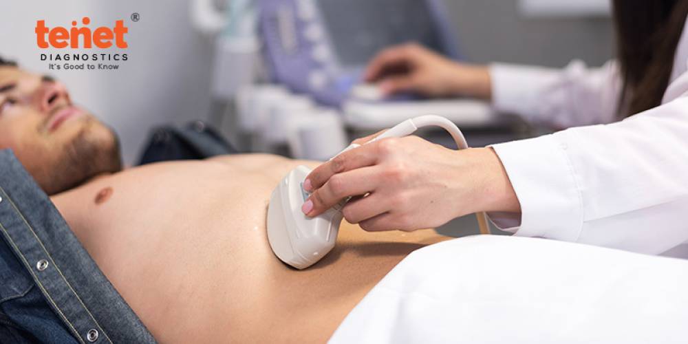
Ultrasound scanning is a diagnostic technique that generates images using high-frequency sound waves. It consists of a tiny transducer (a probe that emits and receives sound) and ultrasonic gel placed directly on the skin. The images are created by moving the transducer along the skin and relaying them to the computer.
Doppler ultrasound, for example, allows the doctor to view and analyze blood flow via the arteries and veins in the abdomen, arms, legs, neck, and brain, as well as throughout the body's organs. The venous Doppler exam is a more focused examination that uses the Doppler ultrasound technique to focus on the prominent veins in the legs (venous doppler test for legs) or arms (venous doppler test for arms).
The Venous Doppler exam detects any obstruction in the veins, commonly a blood clot (or thrombus development) that restricts blood flow. Connect to the best diagnostic center for a venous doppler ultrasound test.
What Is Venous Doppler Test?

Ultrasound imaging is a noninvasive medical diagnostic that aids doctors in diagnosing and treating medical disorders. It is risk-free and painless. It uses sound waves to generate images of the inside of the body. It employs a small probe known as a transducer and gel applied directly to the skin. High-frequency sound waves go from the probe into the body via the gel. The probe gathers the sounds that bounce back. A computer uses these sound waves to generate an image. Ultrasound examinations do not include the use of radiation. Because these ultrasound images are captured in real-time, they can reveal the structure and movement of the body's internal organs. Blood can also be seen flowing via blood arteries in the photos.
Venous ultrasonography visualizes the veins throughout the body. A venous ultrasound examination include a Doppler ultrasound scan. Consult the best diagnostic center near me, Hyderabad, for venous ultrasonography.
Doppler ultrasonography is a type of ultrasound that measures the movement of materials in the body. For example, it allows the doctor to see and evaluate blood flow through the body's arteries and veins.
What Are Some Common Uses of Venous Doppler Test?
The most typical reason for a venous ultrasonography scan is to look for blood clots, particularly in leg veins. Deep vein thrombosis, or DVT, is another name for this illness. These clots may break off and enter the lungs, where they can cause pulmonary embolism, a potentially fatal disease. So, look for the venous doppler test near me to discover an early blood clot in the legs so that treatment can begin to prevent it from spreading to the lung.
1. In addition, a venous ultrasonography scan is performed to:
Identify the source of long-standing leg edema. The valves that ordinarily prevent blood from flowing back to the heart in persons with "varicose veins" may be damaged, and venous ultrasonography can help identify the damaged valves and irregular blood flow.
Assist with the placement of a needle or catheter into a vein. Sonography can assist in locating the vein's exact location and avoiding issues such as bleeding or injury to a neighboring nerve or artery.
The veins in the leg or arm are mapped out so that sections of the vein can be excised and used to bypass a restricted or obstructed blood vessel. One example is using leg veins to surgically bypass constricted heart (coronary) arteries.
Examine a dialysis blood vessel graft if it is not performing as intended; for example, the graft may be constricted or obstructed.
2. Venous ultrasonography scan is used in children to:
Examine a link between an artery and a vein, as seen in congenital vascular malformations (arteriovenous malformations or fistulas) and dialysis fistulas.
Because of the smaller vessel size, if a line is inserted in a vein in the legs or arms, there is a significantly greater likelihood of a clot forming around it (especially in infants and young children).
A clot can form unexpectedly in the arm because of the compression in the vein at the inlet of the chest or in the left leg because of the compression in the vein on the left side by artery in the abdomen in some cases.
Doppler ultrasound enables the doctor to observe and analyze the following:
• Blockages to blood flow (such as clots)
• Tumors and congenital vascular malformations
• Narrowing of vessels
• Increased blood flow (the sign of infection)
• Reduced or no blood flow to various organs, such as the testes or ovary
Procedure Of Venous Doppler Test
.jpg)
A venous Doppler exam is performed at an ultrasound or radiology department of a hospital or a peripheral vascular lab.
There are no prerequisites for this exam. On the other hand, smokers may be urged to stop smoking at least a day before the treatment since smoking constricts blood vessels, which might alter test results.
During the operation, the following can be expected:
Before lying on an examination table or bed, patients are advised to change into a hospital gown for comfort and convenience.
The radiologist will start the venous doppler exam by applying a transparent, water-soluble gel to the skin or the transducer to ensure secure contact with the body and improve sound wave transmission to and from the evaluated area. The radiologist will then move the transducer around the skin in various regions of interest to closely examine the area.
The doctor will look for blood vessels narrowing in the leg's. He may put blood pressure cuffs on various locations on the leg, such as the thigh, calf, and ankle, to compare the blood pressure from the various areas of the leg being evaluated.
The gel will be removed once the imaging process is completed.
The entire venous doppler test procedure usually takes an hour, after which the patient can continue their daily activities.
The results will be submitted to the diagnosing doctor within 2 to 3 hours when the patient is asked to follow up with the doctor. More tests and monitoring may be required if there are some irregularities with the test or if there are worrisome findings. Connect to the best diagnostic center near me to know venous doppler test cost in Hyderabad for the best procedure and accurate results.
Results Of Venous Doppler Test
Normal venous Doppler ultrasonography readings reveal healthy vessels and unimpeded blood flow. Doppler ultrasound abnormal results include the presence of blood clots in veins, closed veins, deep vein thrombosis, and arterial diseases such as arteriosclerosis and arterial occlusion.
Potential Risks and Complications
The venous doppler exam is a noninvasive, painless treatment. There are no hazards; however, there may be discomfort if the transducer passes over a sensitive or tender area. In addition, when the transducer runs over the arteries, it may produce pulse-like noises. The pitch of these noises changes to represent the movement of blood through the veins.
Venous Doppler Test Price
The test price may vary according to the diagnosis labs. But the normal venous doppler test price range is between Rs 2000 and Rs 4000. Connect to the best diagnostic lab near me to know the price of a venous doppler ultrasound test.
Final Words
A venous Doppler is an ultrasound diagnostic test used to assess circulation in the major veins of the legs (or sometimes the arms). This inspection detects vein blockage caused by a blood clot or "thrombus" formation.
Each arm or leg takes about 20 minutes for a venous Doppler ultrasound test. So, Wear clothes that can be easily removed from your waist up (arm imaging) or abdomen down (leg imaging).
The radiologist will compress the veins in your legs or arm to check for blood clots. You may have only minor discomfort depending on the tenderness of your extremities. Then, connect to the best diagnostics in Hyderabad for a venous doppler ultrasound test.